magnetic resonance imaging (mri)
MRIs provide a highly detailed picture of any part of the body and show a high level of detail of the. Open magnetic resonance imaging is required because the member has a documented diagnosis of claustrophobia andor severe anxiety or o When imaging in a physicians office or freestanding imaging center would reasonably be expected to delay care and adversely impact health outcome For code descriptions see the.
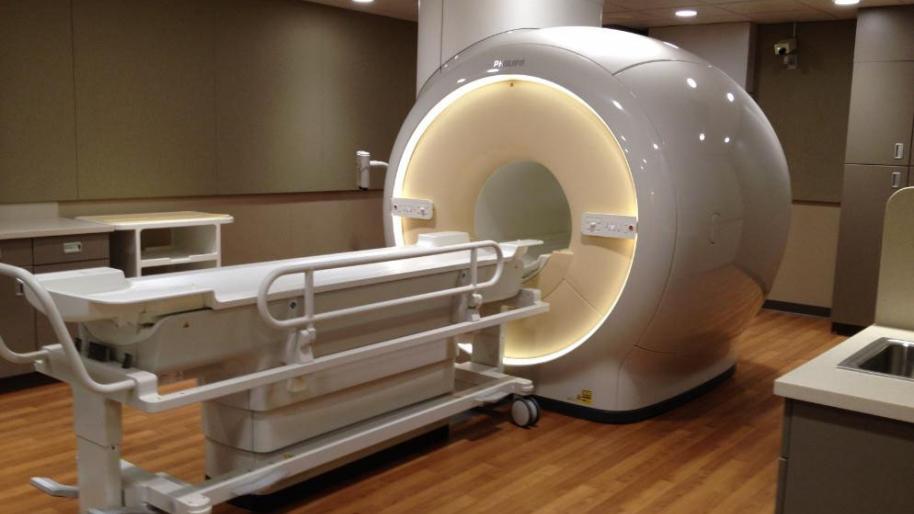
Magnetic Resonance Imaging Mri Michigan Medicine
Unlike X-rays or computed tomography CT scans MRI does not use ionizing radiation.

. Some MRI machines are more open. Magnetic resonance imaging MRI is a test that uses a large magnet radio signals and a computer to make images of organs and tissue in the body. Magnetic Resonance Imaging MRI MR imaging is provided at all Swedish facilities.
Learn more about this Canadian Medical Association accredited program. The didactic courses are offered online three part-time semesters. The Magnetic Resonance Imaging MRI program is a part-time program designed with the working medical radiation sciences professional in mind.
A computer joins all the images and creates a 3D image. Magnetic Resonance Imaging MRI is a non-invasive imaging technology that produces three dimensional detailed anatomical images. Most MRI machines are large tube-shaped magnets.
An MRI scan uses a large strong magnet combined with radio waves to generate multiple cross-section images that are then displayed on a computer. Magnetic Resonance Imaging MRI is a medical imaging procedure for making images of the internal structures of the body. State-of-the-art 3T MRI and cutting-edge molecular imaging are fully integrated as one.
In 2003 Paul C. It may be used to examine the brains functional anatomy determine which parts of the brain are handling critical functions evaluate the effects of stroke or other disease or to guide brain treatment. Magnetic Resonance Imaging MRI Scans.
Some MRI exams include use of injected contrast agent to highlight features in an image and help radiologists diagnose medical conditions. Magnetic resonance imaging MRI is arguably the most sophisticated imaging method used in clinical medicineIn recent years MRI scans have become increasingly common as costs decrease. Magnetic resonance imaging MRI is a noninvasive modality which produces multiplanar and true 3D datasets of subjects in vivo.
Magnetic resonance imaging MRI is a diagnostic procedure that uses a combination of a large magnet radiofrequencies and a computer to produce detailed images of organs and structures within the body. Learn how to help your child prepare. It is often used for disease detection diagnosis and treatment monitoring.
Journal of Magnetic Resonance Imaging JMRI is an international journal devoted to the timely publication of basic and clinical research educational and review articles and other information related to the diagnostic applications of magnetic resonance. Cardiac MR allows very detailed. Magnetic Resonance Imaging MRI is the first international multidisciplinary journal encompassing physical life and clinical science investigations as they relate to the development and use of magnetic resonance imagingMRI is dedicated to both basic research technological innovation and.
MRI scanners create images of the body using a large magnet and radio waves. Modern MRI technology delivers new capabilities to image an ever-changing and growing patient population. The MRI machine is large and tube-shaped.
Crucially it differs from other techniques such as computed tomography CT by producing excellent soft tissue. Lauterbur and Sir Peter Mansfield were awarded the Nobel Prize in Medicine for their discoveries concerning magnetic resonance imaging. No radiation is produced during an MRI exam unlike X-rays.
The magnetic field generated by the MRI can cause damage to hearing aids. In this article we will outline the basic principles behind MRI scans how to orientate and interpret a scan and address some of their advantages and disadvantages. A system that brings a revolution in diagnostic imaging to life.
In our Magnetic Resonance Imaging MRI Graduate Certificate program learn how to you can start a career as an MRI technician or technologist in just eight months. All our scanners are accredited by the American College of Radiology. Magnetic resonance imaging MRI is a medical imaging technique used to produce high quality images of the human body.
In this case the heart is imaged. Magnetic resonance imaging MRI uses a powerful magnetic field radio waves and a computer to produce detailed pictures of the bodys internal structures that are clearer more detailed and more likely in some instances to identify and accurately characterize disease than other imaging methods. MRI scanners use strong magnetic fields and radio waves radiofrequency.
Magnetic resonance imaging MRI is a medical imaging technique that uses a magnetic field and computer-generated radio waves to create detailed images of the organs and tissues in your body. Magnetic Resonance Imaging MRI MRI stands for magnetic resonance imaging and refers to a medical imaging scan that uses a strong magnetic field and radio waves to capture detailed images or pictures of soft tissues bone fat muscles and the internal organs of your body. View full aims scope.
How does an MRI work. Like a computed axial tomography CT scan an MRI photographs the organ several times while a patient lies on a table. The MRI machine is a large cylindrical tube-shaped machine that creates a strong magnetic field around the patient.
Magnetic resonance imaging MRI is a diagnostic exam that uses a combination of a large magnet radio waves and a computer to produce detailed images of organs and structures within the body. Estimating kinetic parameters from dynamic contrast-enhanced t 1-weighted MRI of a. It is based on sophisticated technology that excites and detects the change in the direction of the rotational axis of protons found in the water that makes up living tissues.
Functional MRI fMRI Functional magnetic resonance imaging fMRI measures the small changes in blood flow that occur with brain activity. Take Your Magnetic Resonance Imaging to the Next Level. The wobbling of the atoms is what is recorded by computers and used to reconstruct the images.
MRI uses radio frequency waves and a strong magnetic field rather than x-rays to provide remarkably clear and detailed pictures. This program consists of both didactic and clinical components spread across four semesters. It creates a strong magnetic field around the body.
MRI magnetic resonance imaging is a diagnostic procedure that uses a combination of a large magnet radio waves and a computer to produce detailed images of organs and structures within the body. Our systems are designed to be less claustrophobic for patients. Magnetic Resonance Imaging MRI Cardiac MRI creates images from the resonance of hydrogen atoms when they are polarized to face in one direction and then hit with an electromagnetic pulse to knock them off axis.
Magnetic resonance imaging MRI uses radio waves and magnets to take pictures of organs and structures inside the body by measuring their energy. It is used to evaluate the body for a variety of. Some MRI machines look like narrow tunnels while others.
It achieves high spatial resolution typically of the order of millimeters in the clinical setting. If an injected MRI contrast agent is needed for your imaging an IV catheter will be inserted in your arm by a nurse or technologist. Magnetic resonance imaging or MRI is a noninvasive medical imaging test that produces detailed images of almost every internal structure in the human body including the organs bones muscles and blood vessels.
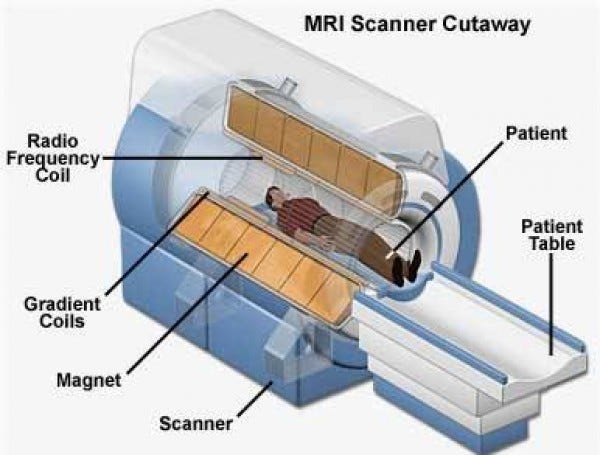
The Story Of Magnetic Resonance Imaging Mri By Deepthi Medium
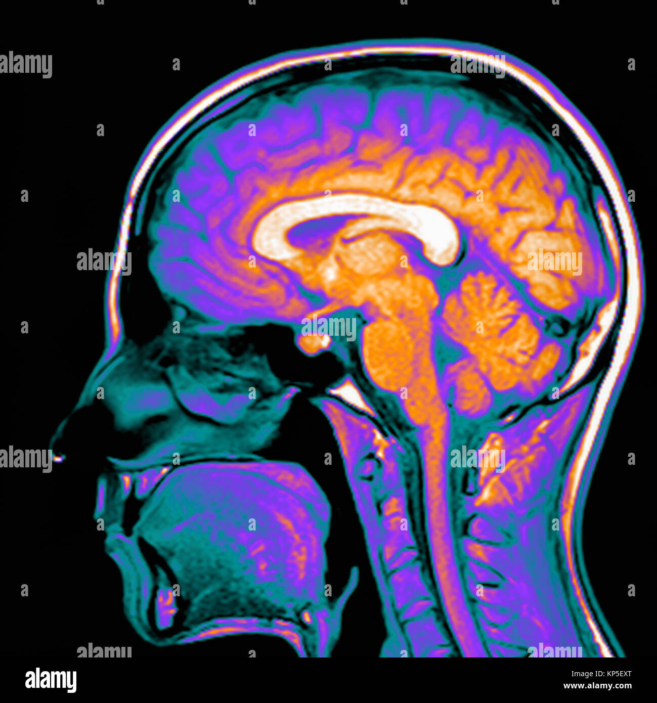
Coloured Magnetic Resonance Imaging Mri Scan Of A Sagittal Section Through A Patient S Head Showing A Healthy Human Brain Stock Photo Alamy

Mri Catholic Health The Right Way To Care
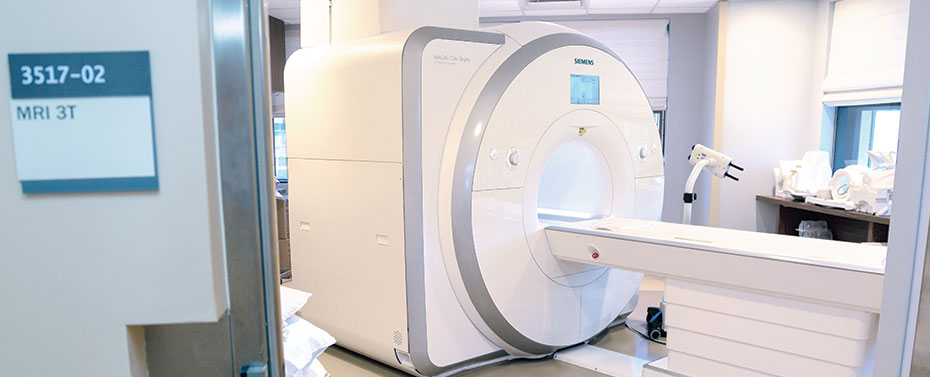
Magnetic Resonance Imaging Mri Englewood Health

Magnetic Resonance Imaging Mri Cohen Medical Centers

Safety Training Tip Magnetic Resonance Imaging Mri Hsi

Magnetic Resonance Imaging Mri Department Of Neurology College Of Medicine University Of Florida

Magnetic Resonance Imaging Mri Piedmont Virginia Community College

Magnetic Resonance Imaging Mri Oklahoma City Ok Spinal Cord Injury Tulsa Ok
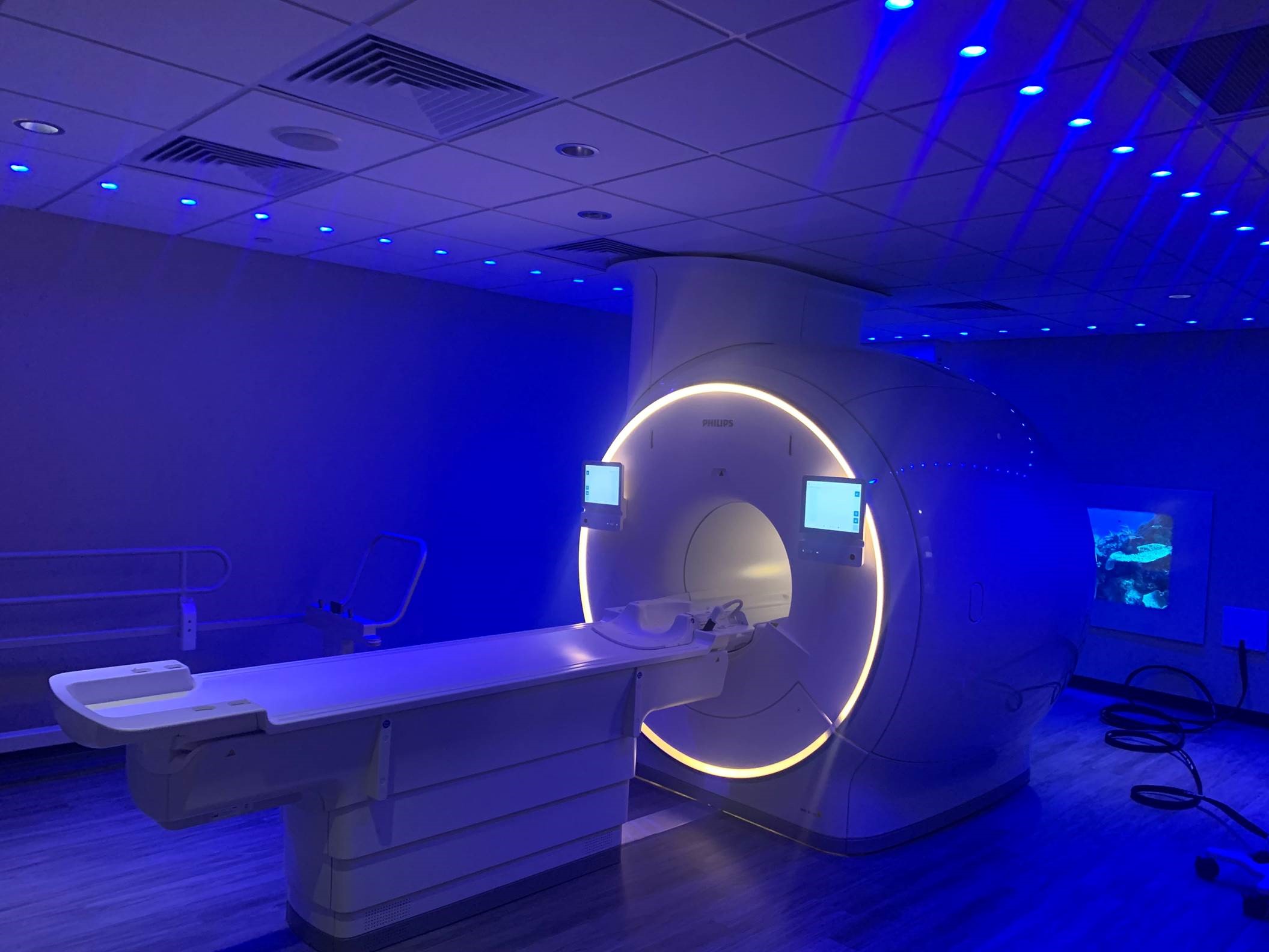
Magnetic Resonance Imaging Mri Carson Tahoe Health
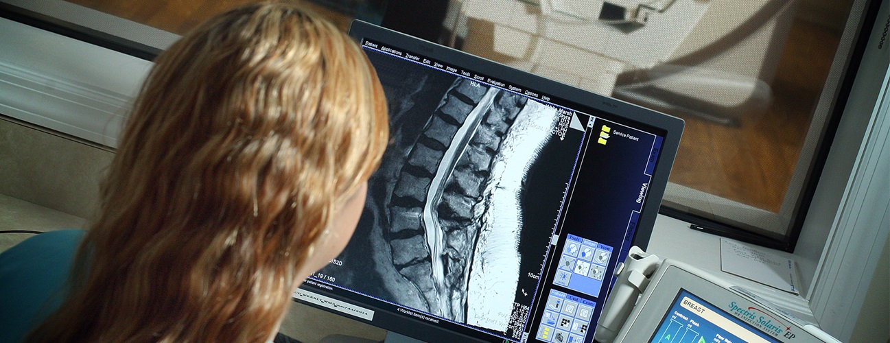
Magnetic Resonance Imaging Mri Johns Hopkins Medicine

Magnetic Resonance Imaging Wikipedia
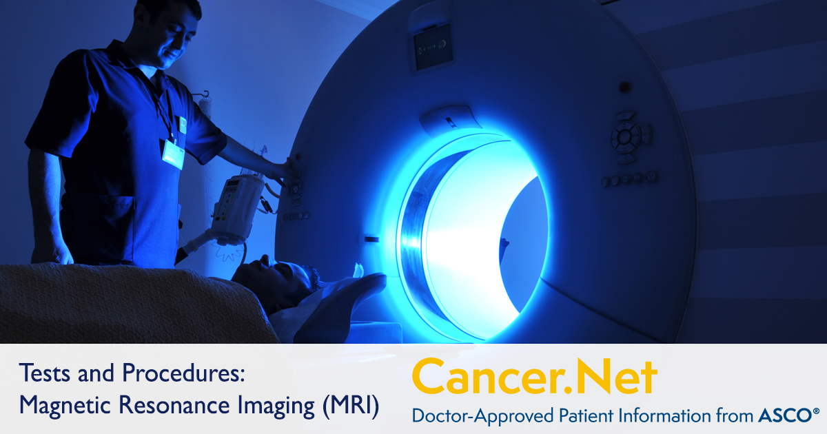
Magnetic Resonance Imaging Mri Cancer Net

Magnetic Resonance Imaging Mri Spartanburg Regional
Magnetic Resonance Imaging Mri

Brain Magnetic Resonance Imaging Mri For Subjects S1 S2 And S4 Brain Download Scientific Diagram

Magnetic Resonance Imaging Mri Special Subjects Merck Manuals Consumer Version

Magnetic Resonance Imaging Mri Of The Body Flat Vector Image
0 Response to "magnetic resonance imaging (mri)"
Post a Comment